Macular Degeneration Test
There is no one macular degeneration test, but rather several ways that your eye care professional can determine if are at risk for developing macular degeneration or if you have macular degeneration.
Visual Acuity Test
A macular degeneration (AMD) eye exam includes several different tests. The first test in a comprehensive eye exam is a Visual Acuity Test.

This test simply tests for the sharpness of your vision. The most common one being the Snellen chart which is the familiar white chart with black letters.
How often you should have a general eye exam depends on your age, if you currently wear prescription glasses and if you have any eye diseases or other health conditions.
The American Academy of Ophthalmology recommends:
"Get a comprehensive eye exam if you have risk factors such as diabetes or a family history of eye disease.
Get a baseline eye exam by age 40, even if your vision seems perfect.
Schedule an eye exam every 1–2 years if you’re age 65 or older."
Be sure to ask your eye doctor if any changes in the retina were found or if any macular drusen were present.
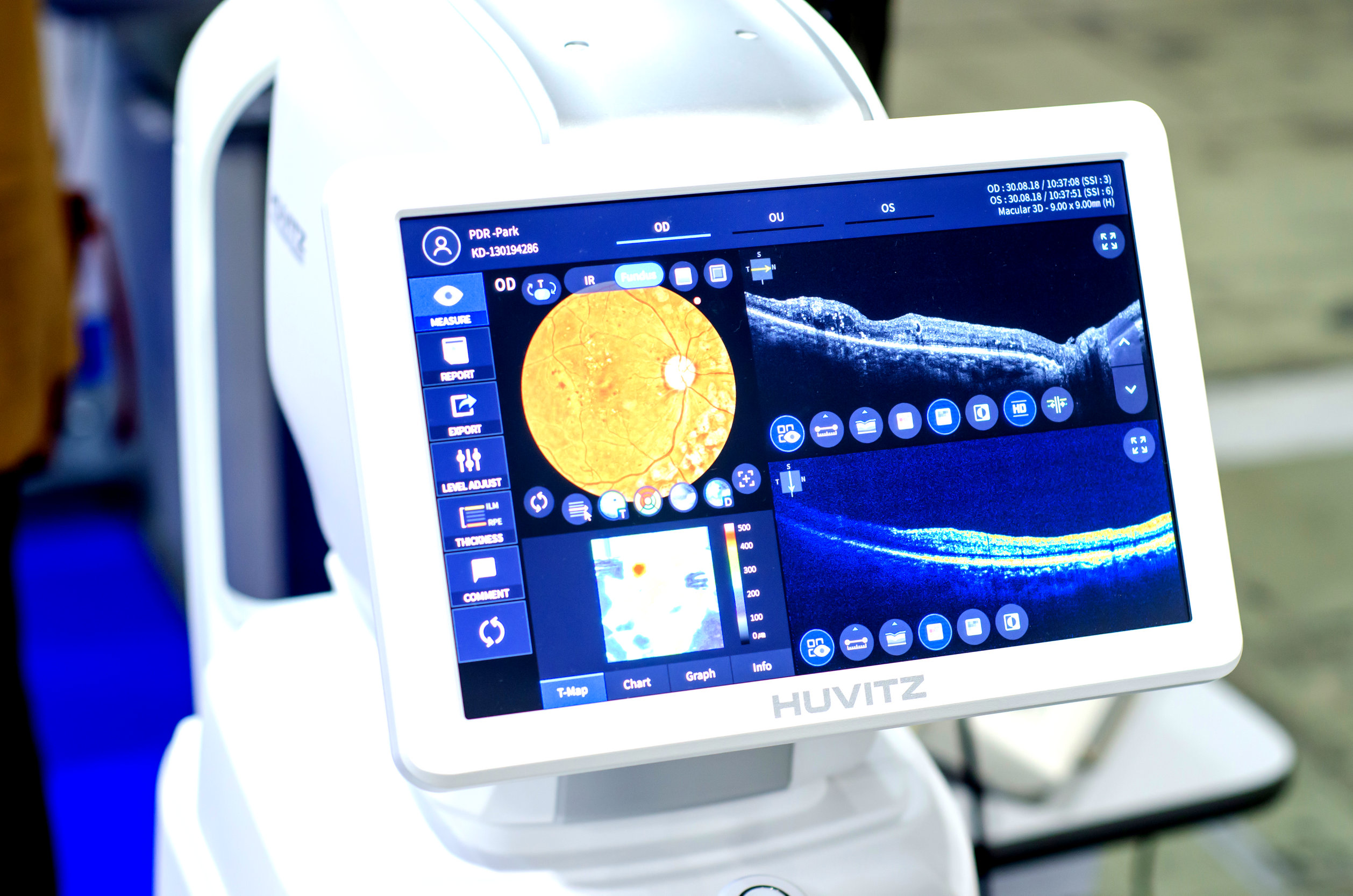
Optical Coherence Tomography
"With optical coherence tomography (OCT), your ophthalmologist can see each of the retina’s distinctive layers. This allows your ophthalmologist to map and measure their thickness. These measurements help with diagnosis. They also provide treatment guidance for glaucoma and diseases of the retina."
What is Optical Coherence Tomography by American Academy of Ophthalmology
Credit for picture from ©lutsenko/123RF.COM
OCT can also be used to monitor one's macular degeneration progress. "It permits to define the location and nature of the changes in the retina and adjacent structures and objectively evaluates the thickness of the retina and surrounding structures.
These capabilities allow detection of newly emerging fluid and/or intraretinal or subretinal tissue and tissue below the retinal pigment epithelium."
Optical Coherence Tomography in Age-related Macular Degeneration
Macular Degeneration Grid or Amsler Grid
The Amsler Grid is a macular degeneration test that checks for any distortion or wavy lines, which are symptoms of AMD. A person with normal vision will see all straight lines.

A person with wet macular degeneration sees something like this illustration - the lines are wavy and distorted. They may also see a "hole" in the middle of the macular degeneration grid.
The doctor is assessing the macula (the center of the retina) when testing your vision using the Amsler Grid. A patient diagnosed with dry or wet macular degeneration should have an Amsler grid at home to monitor their vision. If any changes are noticed, notify your doctor right away.
Find out how to get a free Macular Degeneration Grid and get instructions on how to use this macular degeneration test to check your vision:
Early Detection of AMD
Early macular degeneration testing may soon be offered as part of a regular eye exam.

Rather than looking at the retina or macula, this test actually measures the ability of the eyes to adapt to darkness. My husband has this test done as part of his routine eye exam because of his family history of macular degeneration.
Early Macular Degeneration Detection
Optomap Retinal Exam
Early macular degeneration testing may soon be offered as part of a regular eye exam.

Early macular degeneration testing may soon be offered as part of a regular eye exam. A new technology allows the back of the eye to be seen in a wider view and in a way that captures a digital picture.
The digital picture can be saved and then used for comparison when future exams occur.
At Home Macular Degeneration Test

Once a person has been diagnosed with AMD, their vision needs to be monitored. An at home macular degeneration test, called the Foresee Home Monitoring Program uses a technologically advanced device that can detect vision changes much earlier than one can with an Amsler Grid.
Research has shown that this FDA approved program resulted in earlier detection of symptoms that are associated with the development of wet macular degeneration.
Find out more about how this device works, who is a candidate for home monitoring and how the information get's sent right to your retina doctor:
Retina Examination with Dilated Pupils
Your pupils are dilated so that your eye care specialist can get a fuller view of the retina. Special drops are administered to dilate the retina.
The drops take about 20-40 minutes to work - but it takes about 4 hours to wear off. Bring sunglasses to wear for your drive home and remember that you may not be able to read for a couple of hours.
Here is a picture from the National Eye Institute showing the difference in what an eye professional can see with and without pupil dilation:

After your pupils are dilated, the physician has you sit at a device called a slit lamp.
The slit lamp provides a magnified image of the structures of the eye which helps with a thorough evaluation to detect signs of infection or disease. When used with special lenses, the slit lamp gives the examiner a highly magnified view of the retina.
Fundus Photography
This is when the examiner will look for drusen. A camera can be attached to the slit lamp to take photos of your retina. Drusen are small yellow or off-white deposits that form in the tissue layer underneath the retina.
The appearance of drusen near the macula is one of the most common early signs of macular degeneration. It does not necessarily mean that you have macular degeneration, but it does mean that the eye is at risk for developing AMD. Drusen can be present in the eye for years without affecting your vision.
Drusen that develop away from the macula usually do not indicate the development of macular degeneration. Here is a picture of an eye care professional taking pictures of the retina after dilation. Credit: National Eye Institute

The examiner will also look for other areas of the retina that might appear abnormal such as,
the retina appears raised
bleeding or
fluid behind the retina.
These signs would suggest choroidal neovascularization (CNV)or wet macular degeneration. In these cases, further testing may be necessary.
Macular Pigment Density Test
Measuring the density of the macular pigment provides important information about one's risk of developing age related macular degeneration.

Low Macular Pigment Optical Density (MPOD) is a key risk factor for Age-Related Macular Degeneration (AMD).
QuantifEye is a scientifically-proven instrument that measures Macular Pigment Optical Density (MPOD).
Find out more about this macular degeneration test, who should have it and what to do if you test low for MPOD:
Fluorescein Angiogram
Fluorescein angiography or angiogram evaluates the blood vessels of the retina and helps to make treatment decisions for macular degeneration. It is most useful in diagnosing choroidal neovascularization (CNV) or wet macular degeneration.
A dye is injected into an arm vein which then travels through your blood stream to your eye. You may get a warm feeling or experience a hot flush. This only lasts seconds and then disappears.
Photos of the retina are then taken over a period of about 60 seconds as the dye enters the vessels at the back of your eye. The ophthalmologist can see if the dye leaks from any of the vessels to determine if and where new and fragile blood vessels have developed.
This is how the process works:
1) Pupils are dilated with eye drops
2) Pictures are taken before the dye is given as a baseline
3) An injection of fluorescein dye is given into the vein of the patient's arm
4) They dye travels to the blood vessels in the eye and the retina - takes just seconds
5) Multiple pictures are taken of the retina. The dye gives the blood a glowing appearance so that the location of leaky vessels can be determined. Why is this test given?
This test is ordered if:
1) Your physician suspects that you have wet macular degeneration. It can tell your physician:
√ the pattern of choroidal neovascularization (occult or classic)
√ the boundaries - well defined or poorly defined
√ location of the CNV - subfoveal, extrafoveal or juxtafoveal
√ the size of the lesions
2) To determine the exact location where a leak is to guide any kind of laser treatment.
3) To monitor treatment results for macular degeneration.
There are many ways to incorporate a macular degeneration test at your eye doctor's office to determine if you have AMD or if there is any progression of this retinal disease.
Go from Macular Degeneration Test to WebRN Macular Degeneration Home

√ Prevention of Macular Degeneration?
√ Tips for Daily Living?
√ Food Suggestions for a Macular Degeneration Diet?
√ Ideas on Visual Aids to Maximize your Sight?
If you said "yes" to any of the above, sign up for the monthly Macular Degeneration News.
