Choroidal Neovascularization
Choroidal neovascularization (CNV)is the growth of abnormal blood vessels in the choroid layer of the eye.
CNV can be caused by wet age related macular degeneration and other retinal conditions. There are two types of age related macular degeneration - wet and dry macular degeneration. Wet macular degeneration gets its name from leaking blood vessels in the retina.

Choroid refers to one of the layers of the retina;
neo means new and
vascularization means blood vessels = new blood vessels growing from the choroid layer.
Choroidal neovascularization is a fancy word that simply means there is new blood vessels (neovascularization) growing from the choroid layer of the retina underneath the fovea.
These abnormal blood vessels grow through the Bruch's membrane and into the subretinal space. Wet age related macular degeneration gets its name because fragile, leaky blood vessels grow from this vascular layer and leak into spaces above and below the rods and cones.
When the rods and cones are flooded with this blood and fluid, they are damaged and destroyed.
The result of these photoreceptor cells dying is loss of vision.
Layers of the Retina
The 4 main layers are:
1) The sclera
2) The choroid layer is made of small blood vessels that are responsible for carrying the nutrients to the back of the eye. It is in the middle layer.

3)
Bruch's Membrane
is between the choroid and the Retinal Pigment Epitheliam
(RPE), and acts as a filter between the RPE and choriocapillaries,
keeping them separated.
4) Retinal Pigment Epithelium (RPE) transports metabolic waste from the photoreceptors across Bruch's membrane to the choroid.

The choroid layer contains most of the eye's blood vessels. These new, fragile and abnormal blood vessels grow up through the retinal layers.
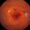
The vessels are very fragile and break easily, causing blood and fluid to pool within the layers of the retina. Here is a picture of wet macular degeneration from the National Eye Institute.
Symptoms of Wet Macular Degeneration
These new and weak blood vessels leak fluid and blood beneath the retina. When this fluid accumulates it raises the normally, smooth surface of the retina which results in these vision changes:
Wet macular degeneration symptoms include:
√ Blurred Vision
√ Distorted Vision

√ Blind or gray spot in the center of vision

The severity of the symptoms depends on:
1) the size of the CNV and
2) its proximity to the macula.
Types of Choroidal Neovascularization
1) Classic Choroidal Neovascularization In classic CNV there is a very rapid leakage of blood and fluid under the retina, causing the surface of the retina to become elevated and uneven. The leakage may even break through some of the layers of the retinal tissue, damaging the retina and leaving blind spots in vision.
2. Occult Choroidal Neovascularization
The blood vessels with this type are "hidden" beneath the fovea and are not readily defined. This type involves a slower blood leak under the retina. Because it is more gradual and there is less fluid, the retina does not become as elevated and uneven as it does with classic CNV.
The vision loss with this type is slower. The vast majority of wet cases are mainly occult or a mix of occult and classic.
Choroidal Neovascularization Treatment
Wet macular degeneration treatment depends on:
√ Size of the bleed
√ Location of the bleed
√ Amount of time that has passed since symptoms first started

The treatment for choroidal neovascularization (CNV) is dependent on where the leaky blood vessels are in relation to the fovea. Bleeding that occurs right under it usually results in worse outcomes... that is because this specific area is made up of cone cells which are photoreceptor cells that are responsible for sharp, detailed vision.
When these cone cells are damaged by blood and fluid, our sharp
and detailed vision is also destroyed. As a result vision is blurred or fuzzy rather than distinct and sharp.
CNV lesions are also classified by where they are located relative to
the fovea (the center of the macula).

1) Sub-Foveal
Subfoveal CNV is a fancy word that simply means there are new blood vessels (neovascularization) growing from the choroid layer (choroidal) of the retina underneath the fovea (subfoveal). This is the most common type of CNV. In fact, almost 90% are the subfoveal type. Subfoveal simply means that the bleeding develops below the fovea.
Treatment options include:
1. Photodynamic Therapy if the CNV is well defined
2. Anti-Vegf Therapy - injections directly into the eye that block the stimulation of new blood vessel growth
2) Juxta-Foveal - lesions in areas other than the center of the fovea
3) Extra-Foveal - lesions that occur in areas of the macula other than the fovea. In this type of wet macular degeneration, CNV develops outside the foveal area.
Go from Choroidal Neovascularization to Wet Macular Degeneration
Go from Choroidal Neovascularization to WebRN Macular Degeneration Home

√ Prevention of Macular Degeneration?
√ Tips for Daily Living?
√ Food Suggestions for a Macular Degeneration Diet?
√ Ideas on Visual Aids to Maximize your Sight?
If you said "yes" to any of the above, sign up for the monthly Macular Degeneration News.
